in vitro studies

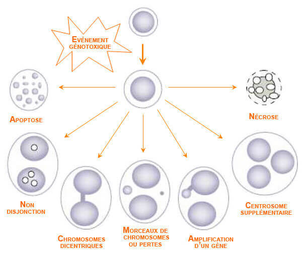
in vitro studies consist in subjecting cells or tissues to low frequency electric and magnetic fields. The objective of in vitro studies is to be able to determine the potential influences of such fields, and to isolate them from other types of influences. However, they also have a major disadvantage: cells or tissues are removed from their natural environment, thereby eliminating the interaction and protection mechanisms otherwise available from the donor organism. Moreover, the fields used are generally stronger than the fields to which the population or workers are exposed. This can result in effects that do not exist with low field values.

It should also be emphasised that a modification that has occured on a cellular level during tests does not mean that the whole organism would experience the same effects.

Note:
DNA damage => possibly genotoxic in humans
DNA damage in vitro => possibly but not necessary DNA damage in vivo

Advantages of in vitro studies

  • Especially important to investigate and identify cellular/molecular working mechanisms:
    • You know exactly what you are doing
    • Your work can be very specific and detailed
      e.g., Investigations of cell division failures by looking at mitotic spindle apparatus or particular DNA studies, ‘omics’,…
  • Fast (fast screening): negative in vitro = negative in vivo
  • Relatively inexpensive
  • Often predictive of a real hazard or risk (e.g., DNA damage)
  • High throughput screening:
    • ex: VITOTOX-test (see further in PubMed)
    • “Omics” (microarray technology, see an example in PubMed)
    • Specific cell lines (lung or skin epithelial cells, white blood cells, hepatocytes,….)

Limitations of in vitro studies

  • Cells are treated outside their normal ‘environment’ (no surrounding tissues, no blood supply, no normal supply of nutrients, …)
  • In vivo exposures can not easily be mimiced
    (Metabolisation can be simulated by addition of specific chemical agents)
    => Enhanced credibility when same effects are also demonstrated in vivo.

In vitro studies - A valid experiment

Importance des points suivants:

  • Travail avec un groupe exposé et un groupe contrôle
  • Double aveugle
  • Mêmes conditions expérimentales
  • Caractéristiques du système d’exposition
  • Lignées cellulaires: tests sur des lignées cellulaires sélectionnées en fonction des objectifs
    • Cellules épithéliales pulmonaires
    • Cellules cérébrales
    • Globules blancs
    • Cellules du foie
    • ….
  • Réplications des études
  • Analyses statistiques

Ces points sont développés dans Etudes in vivo.

Examples of test on cells

Des centaines de tests sont disponibles pour tester les effets d’un agent sur des cellules. Nous présentons ici deux exemples de tests:  le test de cytome et le test de comète. D’autres tests sont également décrits à la page BBEMG: Effets des CEM sur les kératinocytes.

The cytome assay

The cytome assay can be considered as an extended “micronucleus test”; this means that cells are blocked in telophase, just before cell division. In this stage two main nuclei are present. In case of genotoxicity a number of abnormalities are present: micronuclei (broken chromosome fragments or lagging chromosomes are scored in the classical micronucleus test). Other morphological features give additional information: nuclear bridges (dicentric chromosomes), nuclear buds (gene amplification), trinuclear cells (centrosome abnormality). Also numerical chromosome aberrations (e.g., as a result of abnormal nuclear division = non disjuntion) can be scored using specific chromosome probes as well as apoptosis (programmed cell death) and necrosis (cell death).

Etudes in vitro: test de cytome

Source: Fenech M. (2002) Chromosomal biomarkers of genomic instability relevant to cancer.
Drug Discovery Today, 7, 1129-1136.

The single cell gel electrophoresis assay or COMET test

in vitro studies: comet assay

In the comet assay DNA from individual cells is embedded in agarose (gel) on a microscope slide and subjected to electrophoresis (electric current). When DNA is damaged, broken fragments migrate in the gel towards the positive pole. A comet-like structure is formed. The length and the intensitiy of the tail can be measured. Undamaged DNA has no (or very short) tails, the tail lenght is proportional to the damage.

Topics that might interest you ...

Share