Effects on keratinocytes

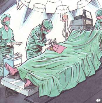
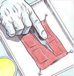
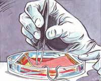
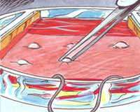
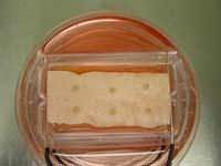
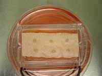
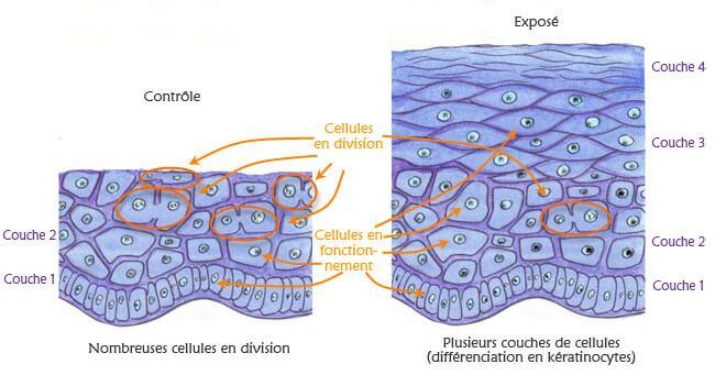
This study intends to analyse the biological effects of extremely low frequency electric and magnetic fields. Keratinocytes, cells of the outermost layer of the human skin (the epidermis), are placed on a piece of decellularized dermis, the inner layer of the human skin. Two platinum electrodes are used to apply the electric signal. Gene activation is compared in exposed cells and in sham exposed cells.

This model of epidermal repair and electric signal provides simplified and well characterized model to study the biological effects of electromagnetic stimulation.

Here is a presentation of the study and the main results.

Dermis preparation

EMF effects on keratinocytes: dermis

The piece of dermis is taken from overweight people during a plastic surgery (abdominoplastia).

EMF effects on keratinocytes: dermis

Exposure to an electric field

EMF effects on keratinocytes: dermis exposure to electric field

The decellularized dermis is placed in a Petri box containing a nutritious medium (in red). Both sides of the dermis are in contact to electrodes.

EMF effects on keratinocytes: dermis exposure to electric field

Keratinocyte samples taken from the same donor, are placed on the dermis. This model mimics the real skin:
Skin = layers of dermis and layers of epidermis cells

Pairs of samples from the same skin donor are prepared for the study. A first one is exposed daily to an electrical field while the other is used as control.

Control

EMF effects on keratinocytes: experiments

Exposed

EMF effects on keratinocytes: experiments

Growth of the epidermis between control and electrically exposed samples
(comparison performed after 14 days of culture)

EMF effects on keratinocytes: experiments

Observation: What is naturally happening is a process of healing (see white area): cells are dividing and the epidermis is growing on the edge of the epidermis samples.However:

  • under sham exposure: larger surface of peripheral cells, which means that they are more actively dividing themselves.
  • under real exposure: larger thickness of the peripheral cells (more layers…)

What is observed under exposure is an enhancement of the cell differentiation (*) at the expense of cell proliferation.

(*) Cell differentiation: This is the process by which a non-specialized cell becomes a specialized cell. In this experiment, it means that cells become faster mature keratinocytes able to play their protective role of the skin.

What’s going on in the peripheral cells (white area)?
With a microscope, cells could be seen as drawn in both illustrations.

Effets des CEM sur les keratinocytes: observation

Illustrations are showing cells in division and other cells (working cells).  According to the aim of this study, we will go further with the latter.

Let us imagine a very powerful microscope that would enable us to make a fine analysis of the inside of a cell and its nucleus. What would we observe?

Effets des CEM sur les keratinocytes: Noyau ADN ARN

DNA

Effets des CEM sur les keratinocytes: Noyau ADN ARN

DNA and RNA

Nucleus contains DNA, a molecule that stores our genetic information. It is organized in sequences called genes: our DNA contains thousands of genes. It is specific for each individual. DNA is a kind of recipe book: it tells how a specific cell will work. However, to be useful, information contained in the recipes needs to reach the cytoplasm, which is the active part of the cells. As DNA is not able to go out the nucleus (the library), another actor enters the scene to send message outside of the nucleus: RNA.

RNA, here represented by the green train, is the transcription of a particular gene. In this example the gene is made up of a succession of six specific bases (elements of the RNA, illustrated here as wagons). The RNA will travel to the cytoplasm (the kitchen of the cell) to be translated into proteins.

More:

When a cell is dividing, DNA is organized into long structures called chromosomes (see figure above). These chromosomes are duplicated before cells divide themselves, which allows both sister cells to receive the whole genetic information.

When a cell is working, genes are transcript into RNA in the nucleus. RNA is a copy of a particular gene that will transfer genetic information from the nucleus to the cytoplasm. Then, it will be transformed into something able to work inside or outside the cells, the proteins. Proteins are the building blocks of the cells and are involved in their functioning.

DNA and RNA are constituted of a succession of only four molecules, called bases.  It is the alphabet of our recipe book. The various combinations of these four “letters” are sufficient to write the whole recipes of our functioning.

Proteins are made up of twenty amino acids (AA). The various combinations of AA give rise to various cooks specialized in various tasks.

What is going on in both samples of cells in this study ? RNAs, transcript in the nucleus, travel to the cytoplasm. It is illustrated below that the amount of RNAs and their origin (the gene transcript) are different under real and sham exposures.

Control

Effets des CEM sur les keratinocytes: Noyau ADN ARN

Exposed

Effets des CEM sur les keratinocytes: Noyau ADN ARN

Unfortunately, the microscope able to see the differences between RNAs does not exist! How scientists could try to identify RNAs and, by the way genes that are activated? It is the mission of the microarray screening. This technique allows analysing gene activation by reporting RNAs quantification. This quantification will allow understanding the cellular mechanisms involved in the cells.

In this study, the aim is to identify the cellular mechanisms triggered by the electromagnetic fields by comparing RNAs from non exposed and exposed cells.

RNA purification

First of all, it is necessary to separate RNA from the other parts of the cells as membrane, organites… This process is called RNA purification. After destruction of cells membranes, RNA are filtered and separated from the other parts of the cells.

Effets des CEM sur les keratinocytes: purification de l'ARN

Before the next step, a labelling molecule is fixed on RNAs. As we will see below, this molecule will allow showing the presence and the amount of specific RNAs on the gene chip. At this moment of the study, researchers have two sets of RNAs: a first one coming from exposed cells and a second one from control cells. Each set of RNA will be separately analysed in order to determine their composition (*).

(*) Note that before being placed in contact with the gene chip, RNAs are transformed into DNAs which is the form recognized by the gene chip. Then we will speak about DNA instead of RNA.

DNA fixation on the gene chip

Both sets of DNAs are placed in contact with a different gene chip. A gene chip is a genius tool for biologists: it is composed of multiple copies of a specific part of well identified genes (e.g. 38 500 for the human species).  These copies are organized on the chip: the same copies are grouped in the same area, like specific stations waiting their trains.

What is happening on the chip when DNAs are placed in contact with the chip? Each DNA recognizes its area and fixes itself on it. Here is a simplified view of a gene chip with only 16 areas of DNA fixation allowing revealing 16 different genes. In reality, this kind of chip is composed of thousands of areas on a surface of less than 2 cm².

Effets des CEM sur les keratinocytes: gene chip

When DNAs are firmly attached to their respective platform, the gene chip is analysed by a scanner able to identify the various areas and to distinguish even the slighter variations in intensity:

  • An area with a more intense coloration is composed of more DNA, which means that the corresponding gene is more activated.
  • On the contrary, when a gene is not or only slightly activated, the area will not show fluorescence.

Both chips are compared in order to analyse RNAs transcript under sham and real exposures.

Gene analysis

After scanning, researchers are informed about DNAs present in both sets and by consequence about gene up or down activated in control and exposed cells. However, let us remember that the gene chip is able to provide information on 38 500 genes! Researchers need to analyse data in order to sort out genes and check their activation level in both sets. The work is huge!

Preliminary results

Up to now, no pathological effect was identified but only acceleration of normal physiological mechanism.

The comparison of the list of significantly up- and down-regulated genes under specific exposure conditions with their respective controls showed the following three up-regulated genes present during all experimental procedures at any sampling time:

  • thioredoxin reductase 1 (TXNRD1),
  • activating transcription factor 3 (ATF3),
  • membrane metallo-endopeptidase (MME).

A comparison of exposed and control groups on day 4 of exposure also showed an up-regulation of Dickkopf Homolog 1 (DKK1) and a down-regulation of microtubule-actin cross-linking factor 1 (MACF1).Regarding the bone formation, we were able to identify one specific protein, BMP2, which is up regulated at day 12 of exposure. This up regulation of BMP2 is able to explain the biological effects observed in all our previous results obtained on cells, embryonic or growing bone tissue and in clinics.The biological functions of these genes confirmed the macroscopic observation: an acceleration of differentiation at the expense of proliferation.

References

Collard J.-F., Hinsenkamp M.
Cellular processes involved in human epidermal cells exposed to extremely low frequency electric fields.
Cell Signal. 27(5):889-98, 2015.

Collard J.-F., Lazar C., Nowé A., Hinsenkamp M.
Statistical validation of the acceleration of the differentiation at the expense of the proliferation in human epidermal cells exposed to extremely low frequency electric fields.
Prog Biophys Mol Biol. 111(1):37-45, 2013.

Collard, J.-F., Mertens, B. & Hinsenkamp, M.
In vitro study of the effects of ELF electric fields on gene expression in human epidermal cells.
Bioelectromagnetics, 32:28-36, 2011.

Hinsenkamp M
Effets des champs électriques et électromagnétiques sur la différenciation cellulaire et leur intérêt en chirurgie orthopédique et traumatologique.
Bulletin et mémoires de l’Académie royale de médecine de Belgique, 166(7-8-9): 307-16, 2011.

Hinsenkamp M., Jercinovic A., Heenen M., De Graef Ch., Wilaert Ch.
Effects of alternating electrical current on keratinocytes in vitro (AC).
Bioelectromagnetics, 18: 250-254, 1997.

Jercinovic A., Hinsenkamp M., Wilaert F., De graef Ch., Heenen M., Goldschmidt D.
Effects of direct constant current on keratinocytes in vitro (DC).
Bioelectroch. Bioener., 39(2): 209-214, 1996.

Topics that might interest you ...

  • Effets des champs sur les os
  • Utilisation des propriétés électromagnétiques – Comment fonctionnent nos appareils électriques? A partir des exemples décrits, nous aurons un aperçu général du fonctionnement d’appareils qui transforment l’énergie électrique en énergie thermique et/ou mécanique ou qui utilisent les propriétés de l’électrostatique et de l’électromagnétisme.
  • Trajet de l’électricité – Comment fonctionne le réseau électrique? Les caractéristiques des réseaux de transport et de distribution et les principaux éléments d’un circuit électrique domestique, dont le câblage monophasé et les méthodes de protection.
Share