Let us imagine an accident during a phase of a soccer match: tibias of both chickens are broken! What happens next?
Effects on bones
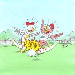
Fortunately for them, these are simple fractures, without bone shifting. No surgery is needed. However, both chickens at least need a plaster. They are advised to rest and walk with crutches for 6 weeks.
Healing callus formation
What happens at the site of the fracture? Fracture healing requires a complex process of cell proliferation and differentiation. It can be divided into 3 major phases:
- The inflammatory phase is characterized by the hematoma formation and physicochemical phenomena that will lead to the migration of the repair cells. These cells will differentiate themselves into specialized cells that will build new bone (cells called osteoblasts) and cartilage (cells called chondroblasts) tissues.
- The reparative phase is characterized by the formation of a callus. It is composed by chondroblasts and osteoblasts that are multiplying in the fracture. The initial callus is quite viscous and do not firmly fix the pieces of bones. After a while, this callus calcifies and the bone becomes more and more rigid.
- The remodeling phase is characterized by the restoration of the normal size, shape and strength of the fractured bone. It may occur after months to years.
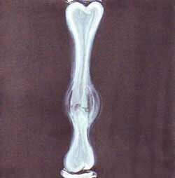
Unfortunately, after this 6-week period, radios are showing an unfriendly image: hyperthrophic pseudarthrosis (*). Pseudarthrosis is a bad prognosis because it means that the fracture is not healing: the callus is staying in its viscous state and is not calcifying.
(*) It is called “hypertrophic” because cell proliferation is very active and bone vascularization is good. Thus, the production of bone increases but the healing tissue does not bridge the fracture. This case of pseudarthrosis, more often due to instability under plaster, is a good candidate for magnetic stimulation treatment.
Why? Various causes are cited for hypertrophic non-union, as for example instability under plaster.
What are the options? How to make bones heal?
Magnetic fields and callus consolidation
Magnetic fields can play a role in callus consolidation! It is not the sole option (for example auto graft, allograft or surgical fixation are other one!) but it can work under specific conditions.
Surprizing? Not necessarily if we go back in the past and travel through history of orthopaedic traumatology (*):
(*) A comprehensive review of the history is available in Hinsenkamp M. Stimulation électromagnétique de l’ostéogénèse et de la consolidation des fractures. Académie Royale de Belgique, Classe des Sciences, Bruxelles, 336 pages, 1994.
As far back as the beginning of the 19th century, electric shocks had been used in the treatment of tibia pseudarthrosis. Techniques have improved and for example in 1861, stimulations involved direct currents applied in the site of the fracture by means of needles but these experiences had no scientific justification.
During the 18th and the 19th centuries, knowledge of electricity developed. As it was the age of the discovery of the electricity and EM field properties, first therapeutic uses of electricity were more “magical” than scientific.
How the idea of using electricity has come to mind and how has the research become more scientific? It can be summarized in three main phases:
1. Observation
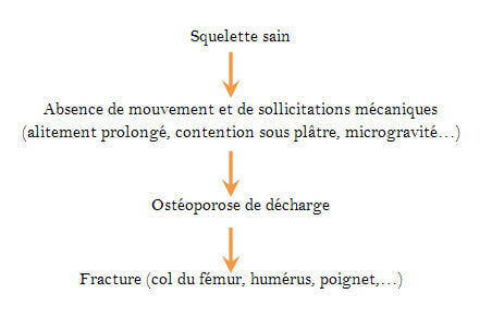
A mechanically solicited bone (for example during a walk) produces bone tissue (osteogenesis). At the opposite, without solicitations bone atrophies (for example, in case of prolonged confinement to bed, plaster cast, microgravity…) inducing a disuse osteoporosis.
2. Experimentations
In parallel, experiments on bones have shown that the repartition of electric charges varies on both sides of a bone according to mechanic deformation. This effect was first similar to a piezo electric effect observed for example in dried conditions on mineral like quartz (see figure below).
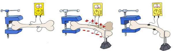
In 1953, scientists observed electrical potential variations at the surface of a bone mechanically deformed. These variations appear during the dynamic phase of the deformation
and cancel out when the deformation is obtained.
However, in living bones (humid conditions), it is essentially the migration of ions during the mechanic deformation that produces a streaming potential, responsible of electrical potential variations at the surface of bones.
3. Therapeutic applications
With these results in mind some authors have emitted the hypothesis that the application of an external magnetic field will lead to electric potential variations at the surface of bones and mimic physiological processes as osteogenesis.
In comparison to previous “historical” therapeutic techniques (e.g. electrical currents applied by mean of needles at the site of the fracture), external magnetic fields are non-invasive: it is simply applied by mean of two coils (see the picture below).
Here below are shown experiments on animals:
Source: Hinsenkamp M. Stimulation électromagnétique de l’ostéogénèse et de la consolidation des fractures. Académie Royale de Belgique, Classe des Sciences, Bruxelles, 336 pages, 1993.
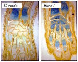
Enhancement of the maturation of the cartilage in mice embryos under in vitro exposure to EM fields (see larger blue areas under stimulation showing an acceleration of the maturation of the cartilage)
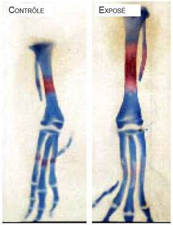
Enhancement of the development of primary ossification centres of chicken bones under in vivo exposure to EM fields (see larger red area under stimulation)
In recent fracture, action of external magnetic fields is characterized by an enhancement of callus rigidity in the first phases of the healing and by the absence of periosteum callus. However, this action does not significantly interfere with the total time of healing of fresh recent fractures.
Happy end
In the present state of the research, even if a slight shortening of the time to healing is observed, it is not sufficient to be applied to humans.
However, modification of the process of healing is observed and further research to precise cellular mechanisms of action of the EMF could improve the treatment of fracture.
What can our chickens expect?
In hypertrophic pseudarthrosis, a large bone cell proliferation is produced usually due to excessive mobilization of the bone fragments and a large bone callus is present but not united. In these conditions, it appears that electromagnetic field could improve healing by acceleration of the maturation of the cartilaginous callus improving its ossification.
At present, it seems that stimulations should start when the callus is already well-developed: callus in its last phases of healing will benefit of magnetic field stimulation by fasting the cartilaginous ossification. Magnetic treatment started too soon could lead to a faster mechanical rigidity of the callus, but less resistant.
In summary
Bone healing is influenced by mechanical factors that external magnetic fields seem to mimic.
A bone stimulated by specific external magnetic fields accelerates bone tissue differentiation (osteogenesis). In other words, the main effect is an acceleration of the cell differentiation. This effect is only seen on sensitive tissues, e.g. in fracture and healing or growing tissues.
Treatments by magnetic stimulations have indications in well-defined cases of hypertrophic pseudarthrosis and ischemical necrosis.
References
Hinsenkamp M., Collard J.-F.
Growth factors in orthopaedic surgery: demineralized bone matrix versus recombinant bone morphogenetic proteins.
International Orthopaedics. 39(1):137-47, 2015.
Hinsenkamp M., Collard J.-F.
Bone Morphogenic Protein–mRNA upregulation after exposure to low frequency electric field. International Orthopaedics.
35(10):1577-81, 2011.
Hinsenkamp M
Effets des champs électriques et électromagnétiques sur la différenciation cellulaire et leur intérêt en chirurgie orthopédique et traumatologique.
Bulletin et mémoires de l’Académie royale de médecine de Belgique, 166(7-8-9): 307-16, 2011.
Hinsenkamp M, Collard J.-F.
Bone Morphogenic Protein–mRNA upregulation after exposure to low frequency electric field.
Int Orthop, 35(10):1577-81, 2011.
Hinsenkamp M.
Influence des facteurs physiques sur la consolidation osseuse.
Bulletin et mémoires de l’Académie Royale de Médecine de Belgique, 151(12): 517-526, 1996.
Hinsenkamp M. Stimulation électromagnétique de l’ostéogénèse et de la consolidation des fractures. Académie Royale de Belgique, Classe des Sciences, Bruxelles, 336 pages, 1994.
Hinsenkamp M.
15 Years Experience in electromagnetic stimulation of bone growth and repair.
J. Jpn. Bioelect. Res. Soc., 8: 1-10, 1994.
Hinsenkamp M.
Clinical studies of the effects of electromagnetic fields on bone tissues.
Cost 244: Biomedical effects of electromagnetic fields. (Ed.: Simunic D.) EU. DG XII: 114-119, 1994.
Hinsenkamp M., Hauzeur J.P., Sintzoff S. Jr.
Preliminary results in electromagnetic field treatment of osteonecrosis.
Bioelectroch. Bioener., 30: 229-235, 1993.
Hinsenkamp M., Heenen M., Dierickx M., Lifschitz L.
Effects of electrical stimulation on wound healing.
Final report contract EC n ° CI1-0348-B, complementary to: CI1-0349-YU: pp. 46, 1993.
Documents & Links
Topics that might interest you ...
- Utilisation des propriétés électromagnétiques – Comment fonctionnent nos appareils électriques? A partir des exemples décrits, nous aurons un aperçu général du fonctionnement d’appareils qui transforment l’énergie électrique en énergie thermique et/ou mécanique ou qui utilisent les propriétés de l’électrostatique et de l’électromagnétisme.
- Trajet de l’électricité – Comment fonctionne le réseau électrique? Les caractéristiques des réseaux de transport et de distribution et les principaux éléments d’un circuit électrique domestique, dont le câblage monophasé et les méthodes de protection.